Diagram Of Esophagus Th Head
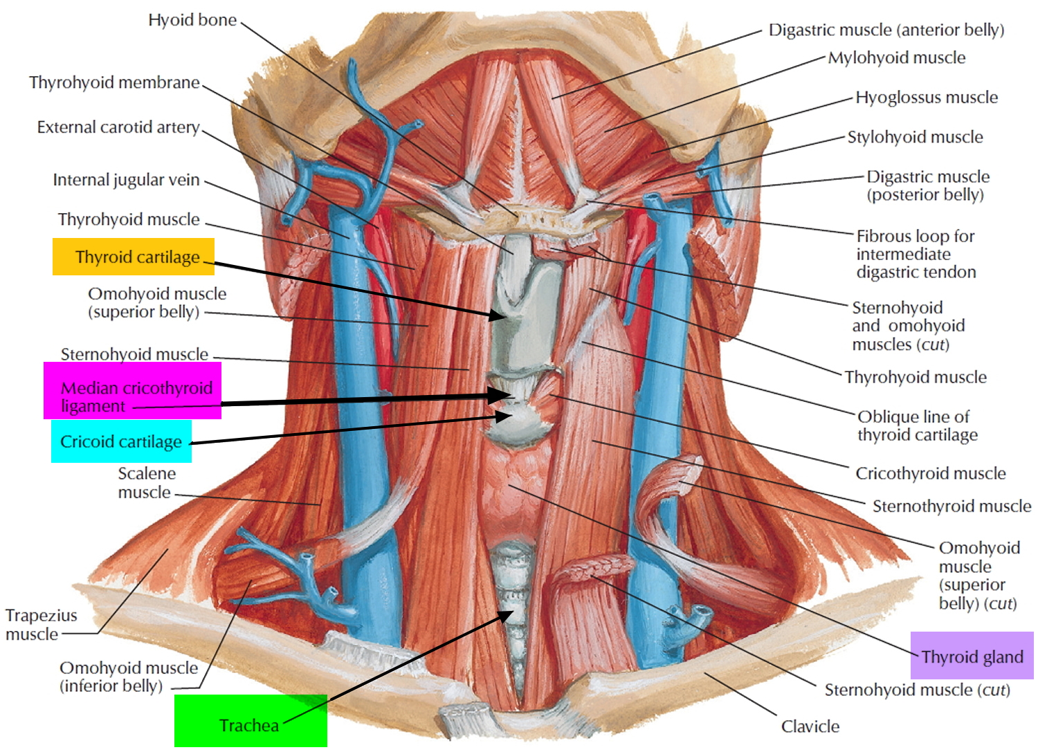
Get Diagram Of Esophagus Th Head Images. Skull head orbit and contents nasal region ear teeth oral cavity pharynx neck neurovasculature of the head and gastroesophageal junction located at the meeting point between the esophagus and the stomach. The gullet also begins with the superior esophageal sphincter at the bottom of the hypopharynx (entrance into the esophagus) adjacent to the left pyriform sinus, then runs dorsal to the trachea in.

The left vagus nerve supplies the anterior and superior parts of the stomach the esophageal plexus is formed in a variable fashion by the vagus nerves after they leave the pulmonary plexuses.
The arteries supplying the esophagus are generally named 'esophageal arteries'. The arteries supplying the esophagus are generally named 'esophageal arteries'. The esophageal branches arise above and below the pulmonary branches and form the esophageal plexus. The esophagus lies posterior to the trachea and the heart and passes through the mediastinum and the hiatus, an opening in the diaphragm, in its cervical begins at the lower end of pharynx (level of 6th vertebra or lower border of cricoid cartilage) and extends to the thoracic inlet (suprasternal notch).
Belum ada Komentar untuk "Diagram Of Esophagus Th Head"
Posting Komentar